Anatomy Of Musckes Sndctendons : Hand Model W Muscles Tendons 3 Part 3b Scientific 1000349 M18 : The muscular system is responsible for the movement of the human body.
byAdmin•
0
Anatomy Of Musckes Sndctendons : Hand Model W Muscles Tendons 3 Part 3b Scientific 1000349 M18 : The muscular system is responsible for the movement of the human body.. A collection of anatomy notes covering the key anatomy concepts that medical students need to learn. The primary function of the knee is to hinge at the lower extremity. The tendons of these muscles pass through a small corridor in the wrist known as the carpal tunnel. Each of these muscles is a discrete organ constructed of skeletal muscle tissue, blood vessels, tendons, and nerves. The anterior and middle scalenes originate from the transverse processes of certain cervical vertebrae and attach to the first rib.
The muscular system consists of the skeletal muscles and their associated structures. Discover the muscle anatomy of every muscle group in the human body. Convergent muscles contain fibers that have a wide origin, but converge in order to attach to a narrow tendon. There's no strict demarcation or dividing line between the tendon and the covering around this muscle but that covering is called is called the epimysium fp my cm and it's really just connective tissue that covers the muscle kind of protects it reduces friction. Muscles of the thorax & abdomen | anatomy model.
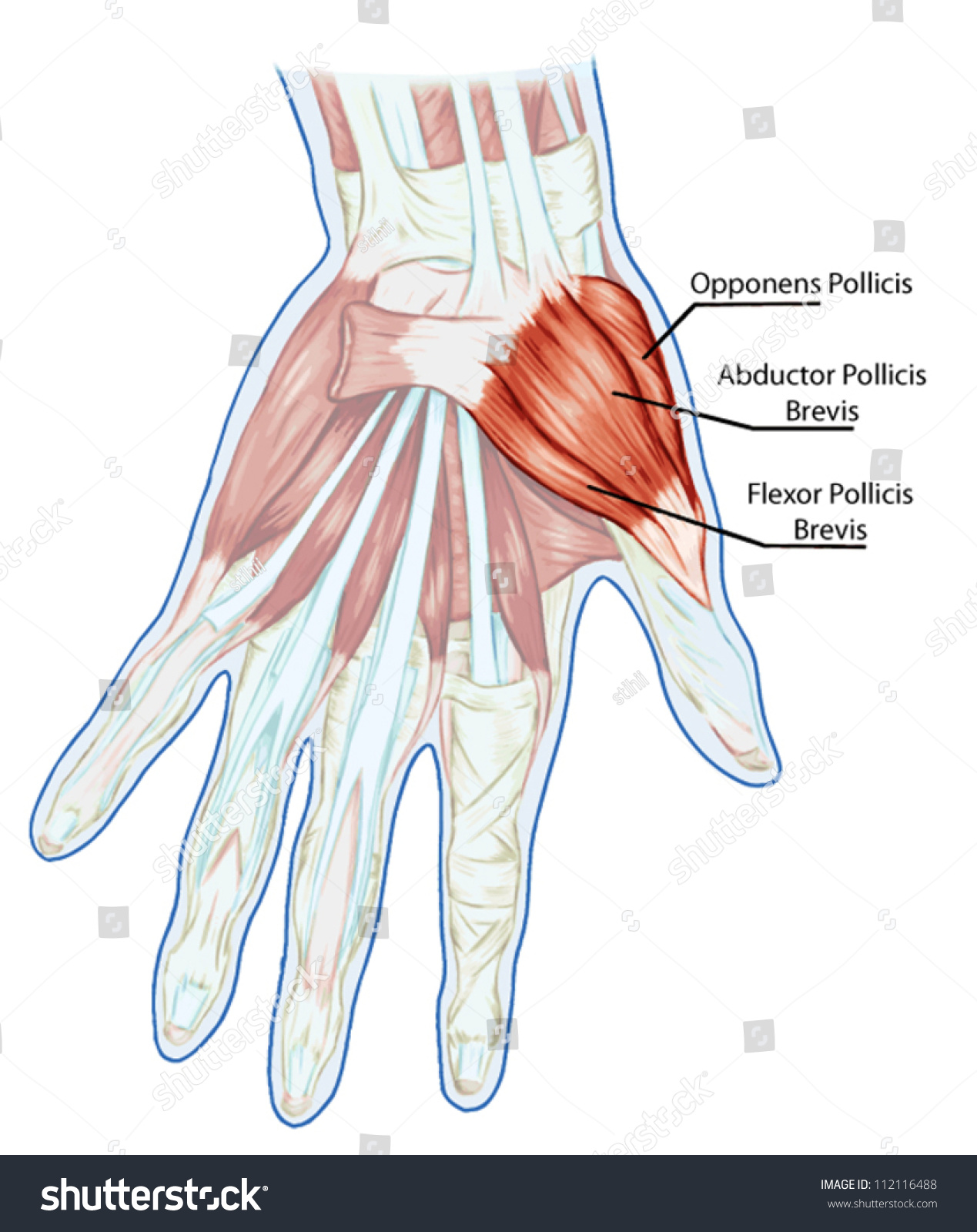
Anatomy Muscular System Hand Palm Muscle Stock Vector Royalty Free 112116488 from image.shutterstock.com Circular skeletal muscles are made up of fibers explore the minute details of the muscular system in complete anatomy with a suite of 3d learning features such as muscle motion, innervation. There are over two dozen gorgeous and painstakingly. Learn about human anatomy muscles with free interactive flashcards. Skeletal muscles allow the body to move and maintain posture; There are around 650 skeletal muscles within the typical human body. Muscle mass accounts for a large majority of the carcass weight of domestic animals. Muscles of the thorax & abdomen | anatomy model. All calf muscle strains result in tearing of some muscle fibers.
Pulling the muscle refers to stretching the calf muscle beyond its limit.
See the pictures and anatomy description of knee joint bones, cartilage, ligaments, muscle and tendons with resources for knee problems & injuries. The primary function of the knee is to hinge at the lower extremity. In the diagrams below, i'll be showing muscle groups in color, with a black line to show the forms that would show through the skin (i also show protruding bones that would do the same). The muscle groups of the upper leg region are the gluteal group. • muscle tissues develop from embryonic cells. Muscle movements, types, and names. There are around 650 skeletal muscles within the typical human body. By contracting, they also aid the venous return of blood to the heart and with age, these components of the musculoskeletal system progressively degenerate, which contributes to frailty and increases the risk of falls and fractures. • the muscular system develops from intra embryonic mesoderm. The muscles of the abdomen may be divided into two groups: A collection of anatomy notes covering the key anatomy concepts that medical students need to learn. The three scalene muscles are found forming the floor of the posterior triangle. As the skeletal muscles pull on bones to cause movements, they also stabilize the joints of the skeleton;
This fascicular organization is common in muscles of the limbs; A calf muscle strain is commonly called a pulled calf muscle. Discover the muscle anatomy of every muscle group in the human body. Pulling the muscle refers to stretching the calf muscle beyond its limit. By contracting, they also aid the venous return of blood to the heart and with age, these components of the musculoskeletal system progressively degenerate, which contributes to frailty and increases the risk of falls and fractures.
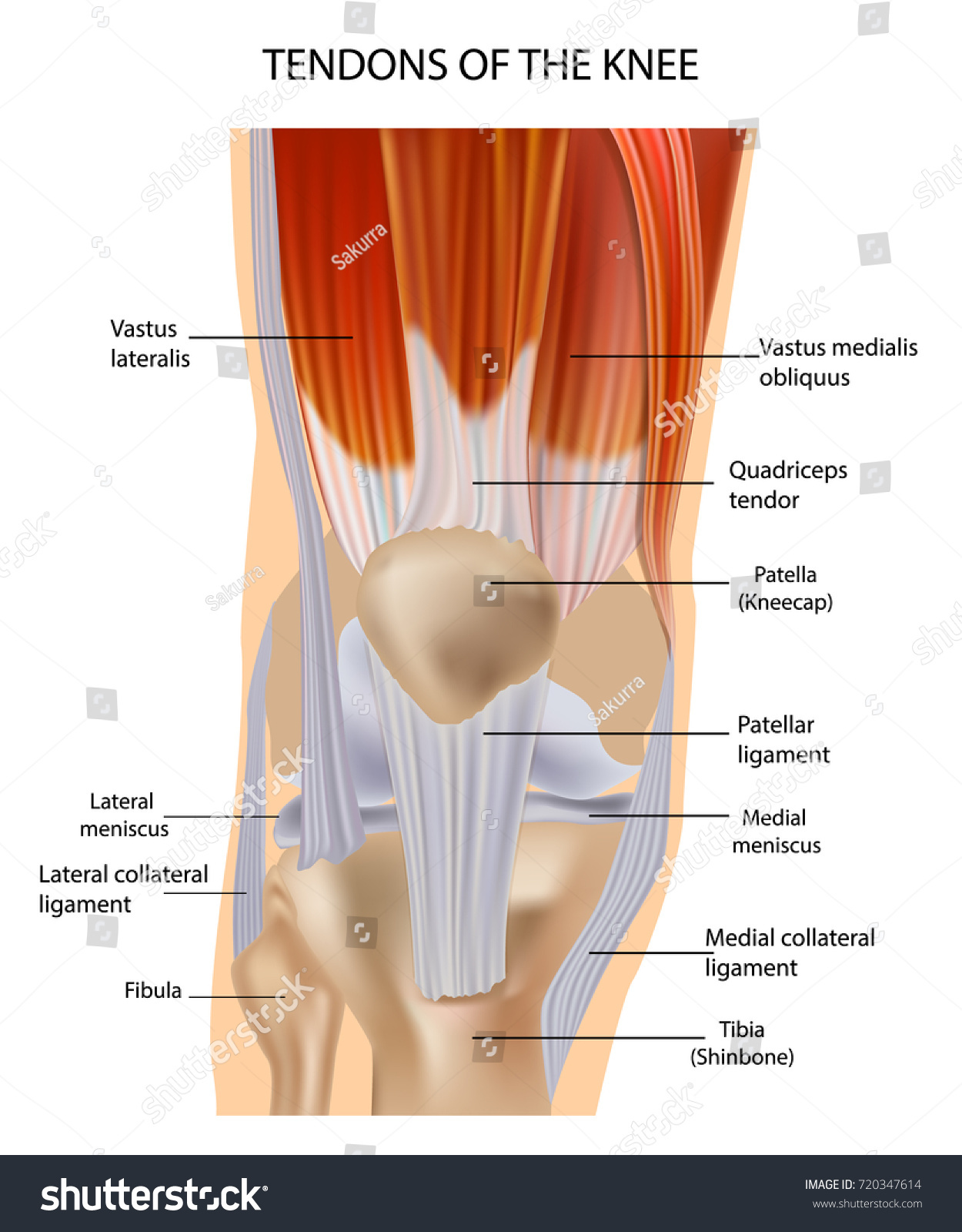
Knee Anatomy from embed.widencdn.net Understanding the structure of a muscle fiber. Skeletal muscles are attached to bones by tendons and can be as long as 30 cm, although they are usually 2 to 3 cm in length. Learn about human anatomy muscles with free interactive flashcards. Microscopic anatomy of skeletal muscle. By contracting, they also aid the venous return of blood to the heart and with age, these components of the musculoskeletal system progressively degenerate, which contributes to frailty and increases the risk of falls and fractures. The muscles of the abdomen may be divided into two groups: Attached to the bones of the skeletal system are about 700 named muscles that make up roughly half of a person's body weight. This article will focus on tongue embryology, origin, insertion, and action of the extrinsic muscles, followed by innervation, blood supply and lymphatic drainage of the tongue.
More serious injuries may result in partial or complete tear of the calf.
Muscle mass accounts for a large majority of the carcass weight of domestic animals. Muscle tendons are extremely important in reinforcing and stabilizing joints. Muscle movements, types, and names. Inflammation of this region caused by repetitive stress or trauma may lead to pain and numbness known as carpal tunnel syndrome. Learning to draw muscles may conjure medical charts in daunting details, but such complexity is unnecessary. Circular skeletal muscles are made up of fibers explore the minute details of the muscular system in complete anatomy with a suite of 3d learning features such as muscle motion, innervation. Microscopic anatomy of skeletal muscle. The muscular system consists of the skeletal muscles and their associated structures. More serious injuries may result in partial or complete tear of the calf. As the skeletal muscles pull on bones to cause movements, they also stabilize the joints of the skeleton; The muscles of the abdomen may be divided into two groups: All calf muscle strains result in tearing of some muscle fibers. Anatomy of the muscular system.
• muscle tissues develop from embryonic cells. Learn about the muscles, tendons, bones, and ligaments that comprise the knee joint anatomy. The primary function of the knee is to hinge at the lower extremity. There's no strict demarcation or dividing line between the tendon and the covering around this muscle but that covering is called is called the epimysium fp my cm and it's really just connective tissue that covers the muscle kind of protects it reduces friction. Attached to the bones of the skeletal system are about 700 named muscles that make up roughly half.
Knee Anatomy Muscles Tendons Muscle Structure Stock Vector Royalty Free 720347614 from image.shutterstock.com Muscle movements, types, and names. Topographically, the muscles in this group are classed along with the lateral torso wall and upper extremity, which is due to their location as well as their genetic development based on their embryological origin. The muscles of the abdomen may be divided into two groups: Related online courses on physioplus. A collection of anatomy notes covering the key anatomy concepts that medical students need to learn. Muscular contraction is necessary for voluntary and involuntary movement of limbs, stabilization of joints, maintaining luminal diameter (in the case of arteries, bowel, etc), and to produce heat. Skeletal muscles allow the body to move and maintain posture; Learn about human anatomy muscles with free interactive flashcards.
There's no strict demarcation or dividing line between the tendon and the covering around this muscle but that covering is called is called the epimysium fp my cm and it's really just connective tissue that covers the muscle kind of protects it reduces friction.
Skeletal muscles allow the body to move and maintain posture; This fascicular organization is common in muscles of the limbs; There are over two dozen gorgeous and painstakingly. The muscular system is responsible for the movement of the human body. Muscular contraction is necessary for voluntary and involuntary movement of limbs, stabilization of joints, maintaining luminal diameter (in the case of arteries, bowel, etc), and to produce heat. Muscle movements, types, and names. In the diagrams below, i'll be showing muscle groups in color, with a black line to show the forms that would show through the skin (i also show protruding bones that would do the same). The anatomy of muscle cells differs from that of other body cells and biologists have applied specific terminology to different parts of these cells. • the muscular system develops from intra embryonic mesoderm. In this section, learn more about the anatomy of the muscles of the neck. The tendons of these muscles pass through a small corridor in the wrist known as the carpal tunnel. Discover the muscle anatomy of every muscle group in the human body. There are around 650 skeletal muscles within the typical human body.